Home » Without Label » Sketch And Label Of A Cross Section Of A Long Bone / The Skeletal System | Biology for Majors II - Continue to label this drawing as you explore the inside of the bone.
Sketch And Label Of A Cross Section Of A Long Bone / The Skeletal System | Biology for Majors II - Continue to label this drawing as you explore the inside of the bone.
Sketch And Label Of A Cross Section Of A Long Bone / The Skeletal System | Biology for Majors II - Continue to label this drawing as you explore the inside of the bone.. A section view is a view used on a drawing to show an area or hidden part of an object by cutting away or removing some of that object. Label the haversian canal, osteocyte (mature bone cell) in lacuna, and canaliculi. The diaphysis of a long bone is composed of bone tissue while the epiphysis is made of 3. The original can be viewed here: This is an online quiz called label the long bone.
A long bone is a bone that has greater length than width. The end of a growing tibia, cut lengthwise*. You need to get 100% to score the 10 points available. The digital cushion sits just behind the pedal bone and above the sensitive frog. Cross section of long bone.
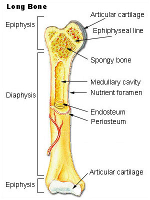
Cross section of tubular bone as appeared in a transmit ... from www.researchgate.net The head of each end of a long bone consists largely of spongy bone and is covered with hyaline cartilage. A long bone is a bone that has greater length than width. The structure of a long bone consists of several sections:. External circumferential lamellae, osteon, central canal, perforating canals, lacuna, canaliculi, concentric lamellae. The diaphysis is the tubular shaft that runs between the proximal and distal ends of the bone. The humerus is the long bone in the upper arm. The digital cushion sits just behind the pedal bone and above the sensitive frog. Looking at a bone in cross section, there are several distinct layered regions that make up a bone.
The structure of a long bone consists of several sections:.
A cross section of a human long bone. In the space provided, draw a longitudinal section of a long bone and label the following parts: (do not copy and paste a picture from the text or internet.) The cut line is called a cutting plane, and can be done in several ways. Plates of cartilage, also known as growth plates which allow the long bones to grow during childhood. Draw a cross section of compact/osteon bone labeling all microscopic structures. A long bone has a shaft and 2 ends. Draw and label a longitudinal section of a long bone. External circumferential lamellae, osteon, central canal, perforating canals, lacuna, canaliculi, concentric lamellae. Forms the larger rounded ends of long bones. Label lines should not cross ; Once we stop growing (between 18. The structure of a long bone allows for the best visualization of all of the parts of a bone ( figure 6.7 ).
The osteocytes are arranged in concentric rings of bone matrix called lamellae (little plates), and their processes run in interconnecting canaliculi. Continue to label this drawing as you explore the inside of the bone. Create a drawing of the bone section in your laboratory journal and label the areas listed above. Diaphysis • shaft of the long bone. External circumferential lamellae, osteon, central canal, perforating canals, lacuna, canaliculi, concentric lamellae.
The Skeletal System | Biology for Majors II from s3-us-west-2.amazonaws.com Make sure learners follow all the criteria for a biological drawing. The humerus is the long bone in the upper arm. The periosteum contains many strong collagen fibers that are used to firmly anchor tendons and muscles to the bone for movement. Bone test anatomy and physiology 12 photos of the bone test anatomy and physiology anatomy and physiology bone lab test, anatomy and physiology bone markings test, anatomy and physiology bone practical test, anatomy and physiology bone tissue test, anatomy and physiology test on bone tissue, bone, anatomy and. Growth in length of a bone occurs at the 4. Draw and label a longitudinal section of a long bone. Area between the diaphysis and epiphysis at both ends of the bone. A cross section of a human long bone.
This is a retouched picture, which means that it has been digitally altered from its original version.
Draw and label a longitudinal section of a long bone. Long bones have a thick outside layer of compact bone and an inner medullary cavity containing bone marrow. Label lines should not cross ; External circumferential lamellae, osteon, central canal, perforating canals, lacuna, canaliculi, concentric lamellae. This is an online quiz called long bone anatomy. This is an online quiz called label the long bone. This slide contained a cross section of a very small bone, and you are looking at the entire thickness of the shaft of the bone. It is located between the elbow joint and the shoulder. A typical long bone shows the gross anatomical characteristics of bone. Proximal epiphysis, distal epiphysis, diaphysis, metaphysis, medullary cavity, epiphyseal line 2. The digital cushion sits just behind the pedal bone and above the sensitive frog. The original can be viewed here: Sketch and label of a cross section of a long bone.
Proximal epiphysis, distal epiphysis, diaphysis, metaphysis, medullary cavity, epiphyseal line 2. End of a long bone. Bone test anatomy and physiology 12 photos of the bone test anatomy and physiology anatomy and physiology bone lab test, anatomy and physiology bone markings test, anatomy and physiology bone practical test, anatomy and physiology bone tissue test, anatomy and physiology test on bone tissue, bone, anatomy and. Cross section of a long bone. Bone remodeling and repair 11.
Kidney-Cross Section | Diagram | Patient from patient.azureedge.net Osteons are oriented parallel to the diaphysis of the long bone. It is located between the elbow joint and the shoulder. Growth in length of a bone occurs at the 4. Cow and human long bones have a similar general structure. The humerus is the long bone in the upper arm. The original can be viewed here: Learners should accurately draw a long bone, resembling that in figure 6.24. Also known as the middle phalanx, the short pastern bone sits on top of the articulating joint of the pedal bone and underneath the long pastern bone.
A long bone has a shaft and 2 ends.
There is a printable worksheet available for download here so you can take the quiz with pen and paper. This is the long central shaft. Proximal epiphysis, distal epiphysis, diaphysis, metaphysis, medullary cavity, epiphyseal line 2. A cross section of a human long bone. Draw and label a longitudinal section of a long bone. The periosteum contains many strong collagen fibers that are used to firmly anchor tendons and muscles to the bone for movement. Related posts of cross section of human bone diagram foot bone anatomy x ray. Once we stop growing (between 18. The axial skeleton runs along the body's midline axis and is made up of. The diaphysis is the tubular shaft that runs between the proximal and distal ends of the bone. External circumferential lamellae, osteon, central canal, perforating canals, lacuna, canaliculi, concentric lamellae. Diaphysis • shaft of the long bone. There is a printable worksheet available for download here so you can take the quiz with pen and paper.